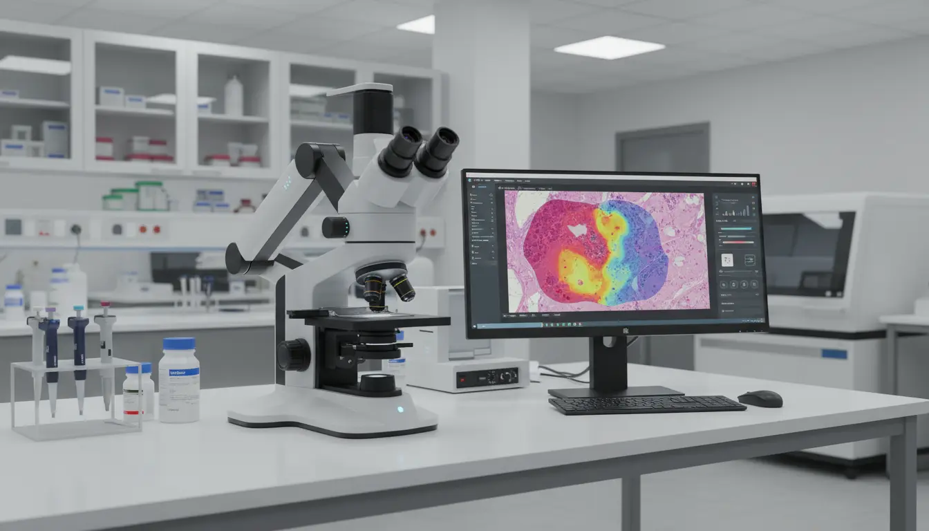

Smart Microscopes: AI‑Driven Automated Diagnosis in Modern Healthcare
Smart microscopes are transforming how clinicians examine tissue and cells. They merge classic optical hardware with powerful artificial intelligence software. Together, they help pathologists diagnose disease faster, more accurately, and more consistently.
Traditional microscopy depends heavily on human expertise and attention. Fatigue, workload, and subtle visual differences can lead to missed patterns. AI‑powered smart microscopes support experts by highlighting suspicious regions automatically. They do not replace specialists but give them a sharper, more reliable second set of eyes.
How AI‑Powered Smart Microscopes Work
Smart microscopes still use lenses, cameras, and illumination like conventional systems. The difference lies in the software layer attached to the imaging pipeline. Images captured by the camera are sent to an AI model in seconds. The model has been trained on thousands or millions of labeled medical images. Using deep learning, the system detects patterns too complex for handcrafted rules. It can recognize cell shapes, tissue structures, and subtle texture differences. The AI assigns probabilities to possible conditions, such as cancer or infection.
The interface then overlays heatmaps, outlines, or color codes on the live image.

Pathologists see both the raw slide and the AI guidance simultaneously. They can zoom, adjust focus, and confirm or reject AI suggestions. Every interaction can also feed future model improvements with proper governance.
Key Benefits for Hospitals and Laboratories
The first major benefit is increased diagnostic speed. AI models process entire slide images in seconds, not minutes. This allows laboratories to handle more samples with the same staff. Patients receive results faster, which is critical for urgent conditions. The second benefit is greater consistency across different experts.
Human readers vary in experience, training, and daily energy levels. Smart microscopes apply the same rules every time a slide appears. They reduce variability and support more standardized reporting practices.
A third benefit is earlier detection of subtle disease signals. Some early lesions are extremely hard to distinguish from normal tissue. AI can notice faint texture or color differences learned from huge datasets. These early flags give specialists a chance to intervene sooner.
Finally, smart microscopes can enhance training and education. Trainees can compare their own slide interpretations with AI feedback. This helps them understand typical errors and refine visual intuition quickly.
Clinical Applications of Smart Microscopes
One prominent application is digital pathology for cancer diagnosis. AI models can grade tumors, count mitotic figures, and measure margins. They assist in classifying breast, prostate, lung, and many other cancers. In hematology, smart microscopes can analyze blood smears automatically. They detect abnormal white cells, red cell morphology, and platelet issues. This helps in diagnosing leukemia, anemia, and various blood disorders.
In microbiology, AI can identify bacteria, parasites, or fungal elements. It can flag suspicious organisms in bodily fluids or tissue samples. This accelerates treatment decisions and supports infection control efforts. Smart microscopes also support telepathology and remote consultation workflows. Slides are digitized, analyzed, and shared securely with distant experts. AI triages cases and highlights the most critical ones for rapid review.
Ensuring Accuracy, Safety, and Regulatory Compliance
Clinical AI tools must meet strict accuracy and safety standards. Developers train models on diverse, well‑curated datasets from multiple sources. They validate performance across different hospitals, scanners, and staining protocols. Regulatory agencies often require rigorous clinical validation studies. These studies compare AI performance with human experts on real cases. Only then can systems be approved for routine diagnostic use.
Even after approval, monitoring remains essential for safe deployment. Hospitals track false positives, false negatives, and user feedback continuously. Models may require periodic retraining to reflect shifting populations and techniques. Data privacy and security are also crucial concerns in medical environments. Smart microscopes must follow encryption, access control, and audit logging standards. Patient data should remain protected across capture, storage, and analysis stages.
Practical Challenges When Implementing Smart Microscopes
Despite their promise, smart microscopes face practical adoption barriers. Initial purchase and integration costs can be significant for smaller labs. Institutions must budget for hardware, software, and network infrastructure upgrades. Workflow integration is another common challenge for clinical teams. AI suggestions need to fit smoothly into existing reporting systems. If interfaces feel clumsy, clinicians may resist regular use.
Trust is a non‑technical but vital adoption factor in healthcare. Pathologists want transparency about how models reach their conclusions. Explainable AI features, such as visual heatmaps, help build that trust. Staff training also matters for safe and effective implementation. Clinicians must understand model strengths, limitations, and failure modes.They should treat AI guidance as support, not unquestioned authority.
Future Trends in AI‑Driven Microscopy and Diagnosis
Future smart microscopes will likely combine multiple data streams. They may integrate genomic data, clinical history, and radiology images. This multimodal context could enable richer, more precise diagnostic insights.
Real‑time decision support will extend beyond hospital walls and labs. Connected devices could assist smaller clinics and resource‑limited regions. Cloud‑based AI analysis may allow high‑quality pathology everywhere with connectivity. Continuous learning systems will refine models using fresh anonymized data. With proper governance, performance can improve without full redevelopment cycles. Feedback loops between clinicians and AI will shape more intuitive tools.
We may also see specialized AI models for rare or complex diseases. These models will help experts handle low‑volume but high‑impact conditions. Smart microscopes will become essential partners across many medical subspecialties.
Building an Ethical and Human‑Centered AI Microscopy Ecosystem
Ethical design must guide every stage of AI microscope development. Teams should examine bias in training data and model outcomes carefully. If certain populations are underrepresented, performance may degrade unfairly.
Involving clinicians, patients, and ethicists helps frame responsible use guidelines. Clear communication should explain how AI affects diagnostic decisions and workflows. Patients deserve transparency about supportive technology used on their samples. Human experts should always remain accountable for final clinical decisions. AI is a tool, not an independent medical authority in the diagnostic process. When used thoughtfully, it amplifies professional judgment rather than replacing it.
Smart microscopes with AI are therefore a strategic investment for healthcare. They support faster, safer, and more consistent diagnostic journeys for patients. With responsible design, they can truly upgrade the quality of global care.